剑麻叶片纤维细胞形态结构差异分析
Analysis of Morphological and Structural Characteristics of Sisal Leaf Fiber Cells
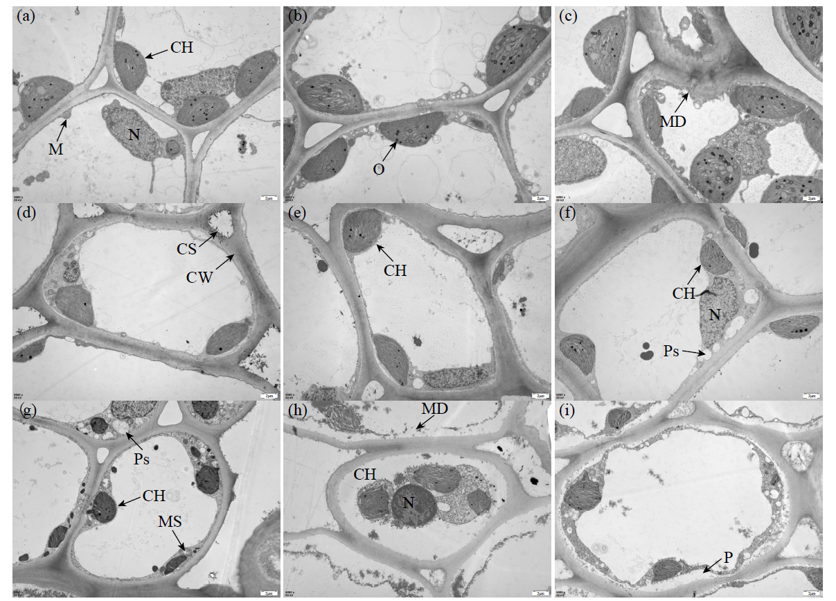
a-c:成熟叶近轴端,d-f:幼年叶近轴端,g-i:稚嫩叶近轴端,CH:叶绿体,O:嗜锇颗粒,Ps:空泡,MS:线粒体肿胀,P:质壁分离;Bar=2μm,加速电压= 80kV
a-c: proximal end of mature leaves, d-f: proximal end of juvenile leaves, g-i: proximal end of infancy leaves, CH: chloroplast, O: osmiophilic, Ps: physalides, MS: mitochondrial swelling, P: plasmolysis; Bar=2μm; accelerating voltage = 80kV