Crops ›› 2022, Vol. 38 ›› Issue (3): 99-103.doi: 10.16035/j.issn.1001-7283.2022.03.014

Previous Articles     Next Articles

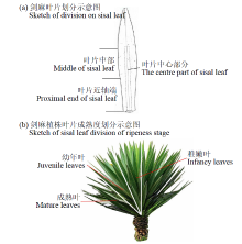
Analysis of Morphological and Structural Characteristics of Sisal Leaf Fiber Cells

Huang Xianya(), Qin Xu, Yang Xiangyan, Li Juxin, Peng Xinyi, Wu Mi, Jin Gang, Chen Tao()   

  1. Guangxi Subtropical Crops Research Institute, Nanning 530001, Guangxi, China
  • Received:2021-08-13 Revised:2021-12-10 Online:2022-06-15 Published:2022-06-20
  • Contact: Chen Tao E-mail:1306475557@qq.com;499481815@qq.com

Abstract:

The purpose of this study was to observe morphological properties of sisal sheet fiber cells and to identify the sampling sites and provide basic experimental references for future studies of sisal sheet fiber cell development. According to the botanical characteristics of leaves and plants, a sampling part was defined, and then used transmission electron microscopy (TEM) observation and analysis of the morphological structural characteristics of the leaf fiber cells of different regions of mature leaves in two growth periods and the proximal end in three maturities degrees, fiber cells in the middle and proximal of old and young sisal were closely related and differ in size. The cells in the center of the leaves were mostly devoid of protoplasts, while protoplasts containing organelles were found at the paraxial end of the leaves. The fibrous cell structure at the proximal end of three maturation stages was further observed. The results showed that various organelles were pushed to the cell edge by vacuoles, and chloroplasts were the most prominent organelles. Among them, the chloroplasts in juvenile and mature leaves were mostly spindle-shaped, the membrane was complete, and a clear lamellar structure and stacking area could be seen. The chloroplasts in the infancy leaves shrinked, the lamellar structure was disordered, the mitochondria swell, vacuoles appear in the cytoplasm, and the phenomenon of increased heterochromatin and separation of the cytoplasmic wall in the nucleus still exists in infancy leaves. From the comprehensive analysis of the image information, it could be concluded that the active site of growth and development of sisal leaf fiber cells is at the paraxial end of the leaf.

Key words: Sisal, Leaf fiber cells, Transmission electron microscopy, Submicroscopic structure

Table 1

The division and maturity definition sampling division of sisal leaves"

序号
Number
依据
Standard
定义
Definition
1 叶片由叶基到叶尖中间1/3部分 叶片中部
2 叶片由叶基到叶尖近叶基1/3部分 叶片近轴端
3 叶片与轴心夹角大于45° 成熟叶(深绿色)
4 叶片与轴心夹角大于30°小于45° 幼年叶(浅绿色)
5 叶片与轴心夹角小于30° 稚嫩叶(黄绿色)

Fig.1

Sketch of sisal leaf division"

Fig.2

Transmission electron microscopy of fiber bundle cells in the proximal end and middle of mature leaves of young and three knife sisal a, c: the proximal end and middle of young sisal leaf, b, d: the proximal end and middle of old sisal leaf, CW: cell wall, CS: cell space, M: mitochondria, N: nucleus, MD: membrane discontinuity, ER: endoplasmic reticulum; Bar=2μm; accelerating voltage = 80kV. The same below"

Fig.3

Transmission electron microscopy of fiber bundle cells at different ripeness stages of sisal leaves a-c: proximal end of mature leaves, d-f: proximal end of juvenile leaves, g-i: proximal end of infancy leaves, CH: chloroplast, O: osmiophilic, Ps: physalides, MS: mitochondrial swelling, P: plasmolysis; Bar=2μm; accelerating voltage = 80kV"

[1] 覃旭, 黄显雅, 杨祥燕, 等. 剑麻麻渣腐熟剂筛选及其腐熟效应研究. 农业研究与应用, 2021, 34(3):48-53.
[2] 李明, 王克荣. 关于亚麻韧皮纤维生长发育阶段划分的探讨. 中国麻作, 1996, 18(1):29.
[3] Li Y, Mai Y W, Ye L. Sisal fiber and its composites:a review of recent developments. Composites Science and Technology, 2000, 60:2037-2055.
doi: 10.1016/S0266-3538(00)00101-9
[4] Li G S, Yin Y J, Li Y, et al. “Steiner trees” between cell walls of sisal. Chinese Science Bulletin, 2009, 54:3220-3224.
doi: 10.1007/s11434-009-0536-1
[5] Li Y, Mai Y W. Interfacial characteristics of sisal fiber and polymeric matrices. The Journal of Adhesion, 2006, 82:527-554.
doi: 10.1080/00218460600713840
[6] 高洸, 刘海敏, 杨之礼. 剑麻纤维的基础研究. 华南理工大学学报(自然科学版), 1992, 20(3):104-114.
[7] 李叶, 黄华平, 邓睿, 等. 不同固定条件对几种植物样品超微结构的影响. 热带作物学报, 2016, 37(11):2100-2105.
[8] 中华人民共和国农业部. 剑麻栽培技术规程:NY/T 222-2004. 北京: 中国农业出版社, 2004.
[9] 刘波. 毛竹发育过程中细胞壁形成的研究. 北京: 中国林业科学研究院, 2008.
[10] 王亮, 邢虎成, 揭雨成. 棉花、苎麻和亚麻纤维细胞发育的激素调控研究进展. 作物研究, 2012, 26(5):602-604.
[11] 王娟, 倪志勇, 吕萌, 等. 棉花纤维伸长期与次生壁增厚期蛋白质组比较. 作物学报, 2010, 36(11):2004-2010.
doi: 10.3724/SP.J.1006.2010.02004
[12] Basra A S, Malik C P. Development of the cotton fiber. International Review of Cytology, 1984, 89:65-113.
[13] Ramsey J C, Berlin J D. Ultrastructural aspects of early stages in cotton fiber elongation. American Journal of Botany, 1976, 63(6):868-876.
doi: 10.1002/j.1537-2197.1976.tb11879.x
[14] 杜雄明, 潘家驹, 汪若海. 棉纤维细胞分化和发育. 棉花学报, 2000, 12(4):212-217.
[15] 甘小洪. 毛竹茎秆纤维细胞的发育生物学研究. 南京:南京林业大学, 2005.
[16] Gan X H, Ding Y L. Developmental anatomy of the fiber in Phyllostachys edulis culm. Bamboo Science and Culture, 2005, 19(1):16-22.
[17] 贺新强, 王幼群, 胡玉熹, 等. 毛竹茎纤维次生壁形成过程的超微结构观察. 植物学报, 2000, 42(10):1003-1008.
[18] 徐明, 任海青, 郭伟, 等. 竹类植物纤维及其细胞超微结构的研究进展. 经济林研究, 2007(4):82-89.
孙焕良, 邝秀明. 苎麻韧皮纤维发育规律研究──I、麻株韧皮纤维细胞发育过程的定位观察. 中国麻作, 1994, 16(3):13-16,51.
[19] 甘小洪, 丁雨龙, 尹增芳. 毛竹茎秆纤维细胞发育过程中ATP酶的超微细胞化学定位研究. 植物研究, 2004, 24(3):357-360,397-398.
No related articles found!
Viewed
Full text


Abstract

Cited

  Shared   
  Discussed   
No Suggested Reading articles found!