
作物杂志,2022, 第3期: 99–103 doi: 10.16035/j.issn.1001-7283.2022.03.014
剑麻叶片纤维细胞形态结构差异分析
黄显雅( ), 覃旭, 杨祥燕, 李菊馨, 彭欣怡, 吴密, 金刚, 陈涛()
- 广西壮族自治区亚热带作物研究所,530001,广西南宁
Analysis of Morphological and Structural Characteristics of Sisal Leaf Fiber Cells
Huang Xianya(), Qin Xu, Yang Xiangyan, Li Juxin, Peng Xinyi, Wu Mi, Jin Gang, Chen Tao()
- Guangxi Subtropical Crops Research Institute, Nanning 530001, Guangxi, China
摘要:
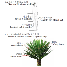
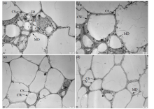
观察分析剑麻叶片纤维细胞形态结构特征,及明确取样部位,为深入研究剑麻纤维细胞发育机制及形态提供基础性和实验性的参考。根据剑麻叶片及植株植物学特征确定取样部位,利用透射电镜(TEM)观察2个生长时期成熟叶片的不同区域和3个成熟度叶近轴端纤维细胞形态结构的差异。由电镜图片可知,剑麻不同生长时期叶片的中部和近轴端纤维细胞紧密相连、大小不一,但叶片中部区域多为不含原生质体的细胞壁结构,而近轴端细胞中存在含有细胞器的原生质体。进一步观察了3个成熟度叶片近轴端的纤维细胞形态,发现叶片纤维细胞中的各种细胞器被液泡挤压到细胞边缘,叶绿体为最突出的细胞器。幼年叶和成熟叶中的叶绿体多呈梭形,外膜完整,可见较清晰的片层结构和垛叠区,稚嫩叶的细胞中叶绿体出现皱缩及片层结构排列较紊乱,线粒体肿胀,胞浆内出现空泡,存在细胞核中异染色质增多和质壁分离的现象。综合分析图像信息推测,剑麻叶片纤维细胞生长发育活跃部位位于叶片近轴端,是观察剑麻纤维细胞结构的理想区域。


| [1] | 覃旭, 黄显雅, 杨祥燕, 等. 剑麻麻渣腐熟剂筛选及其腐熟效应研究. 农业研究与应用, 2021, 34(3):48-53. |
| [2] | 李明, 王克荣. 关于亚麻韧皮纤维生长发育阶段划分的探讨. 中国麻作, 1996, 18(1):29. |
| [3] |
Li Y, Mai Y W, Ye L. Sisal fiber and its composites:a review of recent developments. Composites Science and Technology, 2000, 60:2037-2055.
doi: 10.1016/S0266-3538(00)00101-9 |
| [4] |
Li G S, Yin Y J, Li Y, et al. “Steiner trees” between cell walls of sisal. Chinese Science Bulletin, 2009, 54:3220-3224.
doi: 10.1007/s11434-009-0536-1 |
| [5] |
Li Y, Mai Y W. Interfacial characteristics of sisal fiber and polymeric matrices. The Journal of Adhesion, 2006, 82:527-554.
doi: 10.1080/00218460600713840 |
| [6] | 高洸, 刘海敏, 杨之礼. 剑麻纤维的基础研究. 华南理工大学学报(自然科学版), 1992, 20(3):104-114. |
| [7] | 李叶, 黄华平, 邓睿, 等. 不同固定条件对几种植物样品超微结构的影响. 热带作物学报, 2016, 37(11):2100-2105. |
| [8] | 中华人民共和国农业部. 剑麻栽培技术规程:NY/T 222-2004. 北京: 中国农业出版社, 2004. |
| [9] | 刘波. 毛竹发育过程中细胞壁形成的研究. 北京: 中国林业科学研究院, 2008. |
| [10] | 王亮, 邢虎成, 揭雨成. 棉花、苎麻和亚麻纤维细胞发育的激素调控研究进展. 作物研究, 2012, 26(5):602-604. |
| [11] |
王娟, 倪志勇, 吕萌, 等. 棉花纤维伸长期与次生壁增厚期蛋白质组比较. 作物学报, 2010, 36(11):2004-2010.
doi: 10.3724/SP.J.1006.2010.02004 |
| [12] | Basra A S, Malik C P. Development of the cotton fiber. International Review of Cytology, 1984, 89:65-113. |
| [13] |
Ramsey J C, Berlin J D. Ultrastructural aspects of early stages in cotton fiber elongation. American Journal of Botany, 1976, 63(6):868-876.
doi: 10.1002/j.1537-2197.1976.tb11879.x |
| [14] | 杜雄明, 潘家驹, 汪若海. 棉纤维细胞分化和发育. 棉花学报, 2000, 12(4):212-217. |
| [15] | 甘小洪. 毛竹茎秆纤维细胞的发育生物学研究. 南京:南京林业大学, 2005. |
| [16] | Gan X H, Ding Y L. Developmental anatomy of the fiber in Phyllostachys edulis culm. Bamboo Science and Culture, 2005, 19(1):16-22. |
| [17] | 贺新强, 王幼群, 胡玉熹, 等. 毛竹茎纤维次生壁形成过程的超微结构观察. 植物学报, 2000, 42(10):1003-1008. |
| [18] | 徐明, 任海青, 郭伟, 等. 竹类植物纤维及其细胞超微结构的研究进展. 经济林研究, 2007(4):82-89. |
| 孙焕良, 邝秀明. 苎麻韧皮纤维发育规律研究──I、麻株韧皮纤维细胞发育过程的定位观察. 中国麻作, 1994, 16(3):13-16,51. | |
| [19] | 甘小洪, 丁雨龙, 尹增芳. 毛竹茎秆纤维细胞发育过程中ATP酶的超微细胞化学定位研究. 植物研究, 2004, 24(3):357-360,397-398. |
| No related articles found! |
|
||